PHOTO STORY: A patient with dermatophytosis following tattooing in a cultural context
1 Jul 2025
PHOTO STORY: A patient with dermatophytosis following tattooing in a cultural context
Authors: Swathi N*and Ramesh Bhat M.
Department of Dermatology, Father Muller Medical College, Kankanady, Mangalore, India.
Conflict of interest: None
Key words: Tattoo; dermatophytosis; infection; localized immunodeficiency
Introduction
Tattooing is an ancient practice that holds significant cultural and religious importance in many societies. In India, tattoos are often seen as symbols of religious devotion, social status or personal identity. However, the increasing popularity of tattoos has also led to a rise in associated complications, particularly infections. Although rare, fungal infections such as dermatophytosis is one such complication that can arise from tattooing, especially when proper hygiene and post- procedure care are not followed.1
Case report
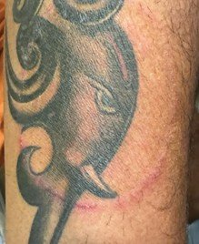
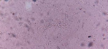
A 23-year-old male, a Hindu by religion, presented to the dermatology clinic with a single pruritic erythematous scaly plaque on his forearm. The lesion had appeared 20 days after he had a tattoo inked in the same area. There were no similar lesions elsewhere or in the family. The tattoo was performed at a local parlour with an intent to honour his religious belief. He also admitted to not following any specific aftercare regimen. On examination, the annular plaque measured approximately 5 cm in diameter, with well-defined borders and central clearing, consistent with a diagnosis of tinea corporis (Fig. 1).


A potassium hydroxide preparation of skin scrapings confirmed the presence of hyphae, indicative of a dermatophyte infection (Fig. 2). The patient was managed with oral itraconazole, 100 mg daily for a period of 2 months in combination with topical antifungals, resulting in complete resolution of the lesion.
Discussion
This patient’s case highlights the intersection of cultural practices and dermatological health. In this instance, the combination of tattooing – a procedure that breaches the skin’s barrier, and the lack of proper post-tattoo care likely facilitated the development of dermatophytosis.
Tattoo-related dermatophytosis, though uncommon, is a recognized complication, developing within 1–3 weeks of skin inoculation and coinciding with the tattoo’s healing period.2 The combination of black ink and micro-trauma from tattoo needles can lead to localized immune disruptions, known as a Ruocco’s immunocompromised district.3 This altered immune state can increase vulnerability to various infections, including dermatophytosis and viral infections such as verrucae and molluscum contagiosum. This is particularly prevalent in settings where hygiene practices are not stringent. The use of non-sterile instruments, improper aftercare and reliance on traditional remedies can all increase the risk of infection.
This patient’s case highlights the need for educating individuals about the potential risks associated with tattooing and the importance of adhering to medical guidelines for wound care.
References
- Cohen PR, Crowley CS, Erickson CP et al. Tinea and tattoo: a man who developed tattoo-associated tinea corporis and a review of dermatophyte and systemic fungal infections occurring within a tattoo. Cureus 2022; 14:e21210.
- Oant¸a˘ A, Irimie M. Tinea on a tattoo. Acta Dermatovenerol Croat 2016; 24:223–4.
- Verma SB. Tinea confined to tattoo sites-an example of Ruocco’s immunocompromised district. Indian Dermatol Online J 2019; 10:739–40.