You are here:
Home
|
What we do
|
Our Foundation
|
CSH Journal
|
French Guiana Tropical Dermatology …
Back to previous page
French Guiana Tropical Dermatology Congress: a digest – Part I
8 Dec 2025
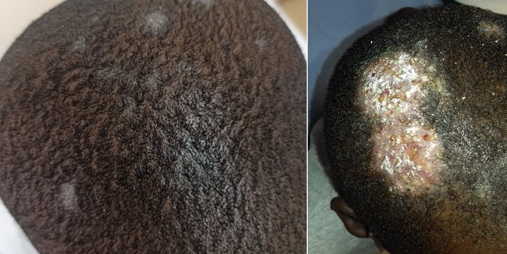
Results
A total of 322 requests for mycological examination of scalp scales were received. The test was positive in 150 cases (46.6%); patients were predominantly male (64.7%) and had an average age of 9 years. Most cases were caused by Trichophyton spp. (141/150, 94%); only 6% (9/150) arose from Microsporum spp.